Extracapsular Ligaments:
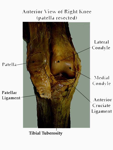
| ligamentum patella: | from inferior border of patella to tibial tuberosity; is the continuation of quadriceps tendon. |
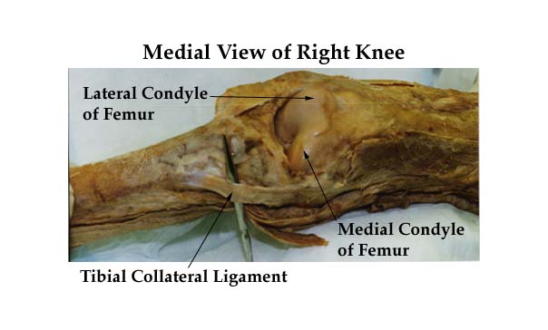
| tibial collateral: | broad flat band attached on medial epicondyle of femur (below adductor tubercle) - runs downward and forward to the medial condyle of the tibia - is crossed by the tendons of sartorius, gracilis and semitendinosus - attaches to meniscus. |
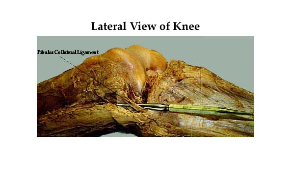
| fibular collateral: | round cord from lateral epicondyle of femur (above groove of popliteus), runs downward and backward to head of fibula - is primarily covered by the tendon of biceps femoris. |
| oblique popliteal: | extension of semimembranosus - attached above the lateral condyle of femur - forms the floor of popliteal fossa and is in contact with popliteal artery. |
| arcuate popliteal: | Y-shaped - from the posterior border of the intercondylar area of tibia and the lateral epicondyle of femur to the area below the head of fibula. |
Intracapsular Ligament and Other Structures: (Netter: 478).
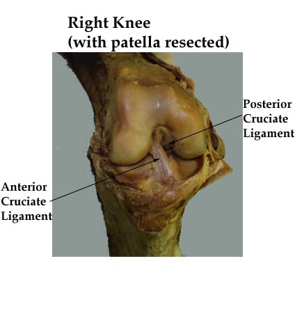
The cruciate ligaments are called cruciate because they cross each other.
| anterior cruciate: | anterior intercondylar area of tibia, runs superiorly, posteriorly and laterally to the posterior part of the medial surface of the lateral femoral condyle. |
| posterior cruciate: | posterior intercondylar area of tibia, runs superiorly, anteriorly and medially to the lateral surface of the medial condyle of the femur. |
Note. The cruciate ligaments:
- are tight both in flexion and extension but are most relaxed at about 30° of flexion, where the collateral ligaments are tight in extension and relaxed in flexion.
- prevent anterior and posterior displacement of tibia where the collateral ligaments prevent abduction/adduction of the knee-joint.
Click to see the drawings below:
|
|
|
|
|
|
|
Click to see the photographs below:
|
|
|
|
|