Articular Surfaces
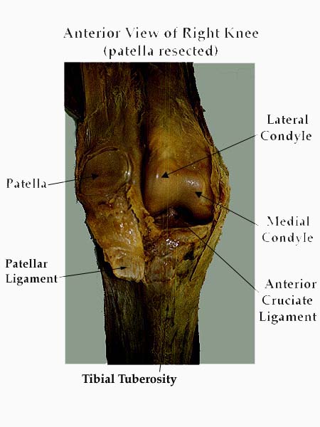
The lower end of the femur (Netter 459) articulates with the upper end of the tibia (Netter 482). NOTE the following features on the femur:
- medial epicondyle
- lateral epicondyle
- medial condyle: oval outline
- lateral condyle: circular outline
- adductor tubercle
- intercondylar fossa
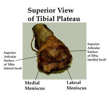
On the tibia: (Netter: 483)
- medial condyle: oval shape
- lateral condyle: circular shape
- medial and lateral intercondylar tubercle
- intercondylar fossa
- tibial tuberosity
- Gerdy's tubercle
- oblique line
- medial and lateral articular facets
Both articular surfaces of the tibia have a corresponding meniscus (a lateral and a medial meniscus). (See later)
The patella is the largest sesamoid bone of the body. A vertical ridge divide the articular surface into a large lateral and a small medial articular surfaces. It is triangular in shape (apex lies inferiorly). It gives attachment for the quadriceps muscles. The patella is thought to increase the pull of the quadriceps.
Click to see the drawings below:
|
|
|
|
Click to see the photographs below:
|
|
|